Li Zhang1,
Xin-Xia Yue2,
Lei Zhang3,
Jin-Fang Zhao4,
Yi-min Chen4,
Zhi-jian Cao5,
Yong-lin Lin4 ![]()
For correspondence:- Yong-lin Lin Email: liuyonglin133494@163.com Tel:+8657187070008
Received: 24 December 2015 Accepted: 4 August 2016 Published: 30 September 2016
Citation: Zhang L, Yue X, Zhang L, Zhao J, Chen Y, Cao Z, et al. Anti-osteoporosis effect of Cistanche deserticola Ma extract in ovariectomized rats. Trop J Pharm Res 2016; 15(9):1929-1933 doi: 10.4314/tjpr.v15i9.17
© 2016 The authors.
This is an Open Access article that uses a funding model which does not charge readers or their institutions for access and distributed under the terms of the Creative Commons Attribution License (http://creativecommons.org/licenses/by/4.0) and the Budapest Open Access Initiative (http://www.budapestopenaccessinitiative.org/read), which permit unrestricted use, distribution, and reproduction in any medium, provided the original work is properly credited..
Purpose: To investigate the therapeutic effects of Cistanche deserticola Ma. extract (CDME) on ovariectomy-induced osteoporosis in rats.
Methods: Female Sprague-Dawley rats were randomly assigned to a control group and five ovariectomy (OVX) subgroups, that is, OVX with vehicle (OVX), OVX with 17ß-estradiol (E2, 25 µg/kg/day), and OVX with CDME doses (40, 80, or 160 mg/kg/day). Daily oral administration of E2 or CDME started 4 weeks after OVX and lasted for 16 weeks. Bone mineral density (BMD) of L4 vertebrae and right femur of rats was estimated, The length of each femur was measured, and biochemical analysis of serum and urine specimens were performed.
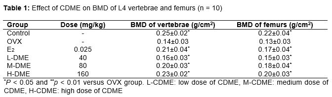
Results: CDME dose-dependently inhibited the reduction in BMD of L4 vertebrae (0.23 ± 0.02 g/cm3, p < 0.05) and femurs (0.20 ± 0.03 g/cm3, p < 0.05) caused by OVX and prevented the deterioration of trabecular microarchitecture (p < 0.05), which were accompanied by a significant decrease in skeletal remodeling (p < 0.05) as evidenced by the lower levels of bone turnover markers.
Conclusion: This study indicates that CDME prevents OVX-induced osteoporosis in rats, and could be used for treating osteoporosis in elderly women.
Introduction
Osteoporosis is the most common bone disease and is characterized by low bone mass, microarchitectural deterioration of bone tissue, and subsequent bone fragility with susceptibility to fracture [1]. Bone fracture risk typically increases in the hip, vertebral, and distal forearm bones. These fractures are not only painful but also disabling, leading to the need for nursing home care and increased mortality when compared to age matched populations [2]. Because of the high morbidity and mortality associated with osteoporotic fractures, treatment of osteoporosis prioritizes fracture prevention [3]. Hormone deficiency is known to impair cancellous metaphyseal bone and reduce bone mineral density in humans and animals; therefore, the estrogen deficiency in postmenopausal women has been regarded as a critical cause of this population’s susceptibility to osteoporosis [4]. Osteoporosis is twice as common in women as in men [5], and approximately one in three old women experience osteoporotic fracture in her lifetime [6,7].
Clinically, hormone replacement therapy (HRT) has been a popular therapeutic strategy designed for post-menopausal osteoporosis [8,9]. However, the long-term application of HRT has potential malignant effects on reproductive tissues [10]. Vitamin D and calcium are components of bone renewal, but supplementation has limited and inconsistent effectiveness and is often used in combination with other treatments [11].
Cistanche deserticola Ma. has been widely used as a kidney tonifying and anti-osteoporosis herb for treating osteoporosis for thousands of years in China [12-14]. Therefore, this study was performed to evaluate the effect of CDME on osteoporosis in rats.
Methods
Preparation of Cistanche deserticola Ma. extract
The herbal samples of Cistanche deserticola Ma. were collected from Bozhou City, Anhui Province in China in September 2015. Taxonomic identification of the plant was performed by Professor Ping He of Zhejiang Chinese Medical University, China. A voucher specimen (no. CDME 201510012) was deposited in Zhejiang Chinese Medical University, China for future reference.
One batch of herbal samples of Cistanche deserticola Ma. was dried in an oven at 80 ℃. Aqueous extract of CDME was obtained by steeping the dried Cistanche deserticola Ma. in water at 70 oC three times for one hour each. Then the extracted fluid was dried in an oven and freeze-dried to obtain the last extract. One gram powder was equivalent to about 1.8 g crude samples. The yield was 55.67 %.
Animals and treatments
Female Sprague-Dawley rats (wt. 200 ± 20 g) were provided by the Experimental Animal Center of Zhejiang Province (Certificate no. SYXK 2003-0004). The animals had free access to feed and water, and were allowed to acclimatize for at least one week before use. The rat experiment was approved by the Animal Care and Use Committee of Zhejiang Chinese Medical University (approval ref no. 20110506) and was carried out in compliance with Directive 2010/63/EU on the handling of animals used for scientific purposes [20].
Sixty rats were randomly divided into six groups of ten individuals: a control group and five ovariectomy (OVX) subgroups, that is, OVX with vehicle (OVX), OVX with 17ß-estradiol (E2, 25 g/kg/day), and OVX with CDME (40, 80, or 160 mg/kg/day).
Bone mineral density (BMD) measurement
The BMD of the L4 vertebrae and right femurs was estimated using dual-energy x-ray absorptiometry scanning with small animal measurement. The measurements were expressed as grams of mineral contents per cm2 of surface area. Scans were performed by the same blinded technician.
Three-point bending test
Before mechanical testing, the left femurs were slowly thawed at room temperature. The length of each femur (distance from the intermalleolar to the intercondylar region) was measured with a micrometer, and the center of the diaphysis was determined.
Biochemical analysis of serum and urine specimens
The levels of serum alkaline phosphatase (ALP), urinary calcium (U-Ca), urinary phosphorus (U-P), and urinary creatinine (Cr) were measured on an automatic analyzer using a diagnostic reagent kit. Serum osteocalcin (OC) concentration was determined using a rat OC ELISA kit.
Statistical analysis
The data are expressed as the mean ± standard deviation (SD). Statistical analysis was performed using one-way ANOVA combined with Bonferroni’s multiple comparison test using SPSS 16.0. Differences were considered statistically significant at p < 0.05.
Results
BMD of L4 vertebrae and femur
demonstrate that OVX significantly decreased the BMD in the L4 vertebrae and femurs compared to control group (both p < 0.05). Compared with the OVX group, CDME treatment obviously prevented the BMD decrease in OVX-induced L4 vertebrae and femurs (p < 0.05) in a dose-dependent manner.
Mechanical characteristics of femur
revealed the results of the femur mechanical testing. Compared with the control group, 16 weeks of estrogen deficiency significantly decreased the maximum load and maximum stress (both p < 0.05). Higher dosage of CDME treatment (80 or 160 mg/kg/day) markedly decreased these parameters (both p < 0.05). E2 also increased the biomechanical properties, which were significantly higher than those of OVX group (all p < 0.05).
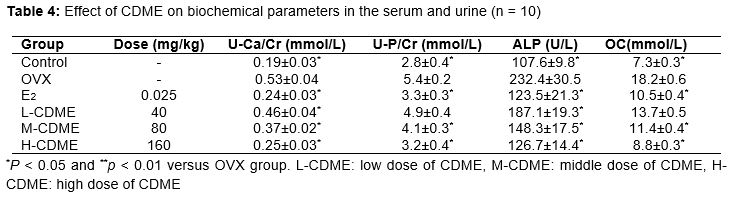
Biochemical profile of serum and urine specimens
The effects of CDME on biochemical parameters in the serum and urine of OVX rats sees . Compared with control group, the levels of U-Ca/Cr, U-P/Cr, ALP, and OC were significantly increased in the OVX group (all p < 0.05). All three CDME doses significantly prevented the increases in U-Ca/Cr and ALP levels (all p < 0.05) in a dose-dependent manner. Higher dosage of CDME treatment (80 or 160 mg/kg/day) significantly prevented the increases in U-P/Cr and OC levels (both p < 0.05).
Discussion
The aim of this study was to determine the potential effects of Cistanche deserticola Ma. extract (CDME) in osteoporosis therapy. Our results suggest that CDME could prevent bone loss. This study revealed that CDME significantly improved bone mass, bone strength, bone microarchitecture, and bone turn-over in OVX-induced osteoporotic rats, which was similar to that of E2. These results suggest the potential role of CDME as a natural alternative for postmenopausal osteoporosis management.
Bone remodeling is the process that mediates changes in the traits that influence bone strength. Any interruption in bone remodeling, such as menopause, will disturb the balance between formation and resorption and cause bone mass loss [16]. Therefore, we used OVX rats as an animal model for human osteoporosis. It has been reported that statistically significant bone loss can be seen after 30 days of treatment [17], so treatment was initiated 4 weeks after OVX. Consistent with other studies, OVX caused significantly higher body weights in our present study, which may be attributed to fat deposition caused by the lack of estrogen. Previous studies suggest that estrogen plays an important role in stimulating the differentiation of progenitor cells through the osteoblast lineage but not the adipocyte lineage [18].
Decreased BMD is one of the major factors that jeopardizing bone strength, resulting in increased susceptibility to fractures [19]. Thus, BMD measurement can best predict fracture risk. Results in the present study showed that OVX reduced BMD in the right femurs and L4 vertebrae, which are rich in trabecular bone, while treatment with CDME dose-dependently prevented the decreases in BMD. Although BMD is among the strongest predictors of facture resistance, both empirical observations and theoretical analyses show that the biomechanical properties of bone and trabecular microarchitecture influenced trabecular bone strength as well [20].
The measurement of bone markers plays a role in osteoporosis diagnosis and treatment [21]. Bone mass loss, as evidenced by enhanced levels of ALP, OC, U-Ca/Cr, and U-P/Cr, indicated upregulation of bone turnover by OVX. The bone turnover markers above were dose-dependently reversed by CDME, indicating a reduction in bone turnover rate after treatment of CDME. This study suggests that CDME is effective for treating osteoporosis in menopausal women.
Conclusion
The findings of this study indicate that CDME prevents OVX-induced osteoporosis in rats, and could be effective for treating osteoporosis in elderly women.
References
Archives


News Updates